Karen Titus
March 2017—The language of blood banking experts, as they talk about irradiators, transfers easily to a car dealership. How reliable are the newer models? Are you willing to replace it every 10 years or so? Do you keep running it until it dies? What parts are likely to burn out? What will repairs run?
And then the word “terrorism” pops up.
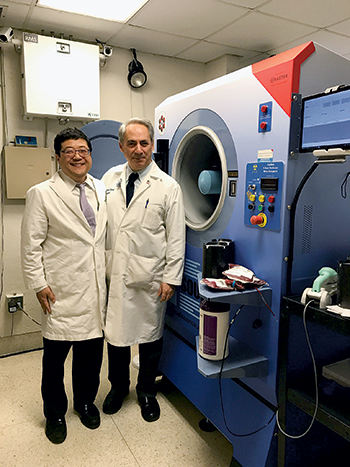
Dr. Jeffrey Jhang (left) and Dr. Jacob Kamen at Mount Sinai Hospital, where the Rad Source RS3400 x-ray irradiator (at right) was installed in January. “We were training, validating, and using the machine in February, and we went live on March 1,” Dr. Jhang says.
Since the Sept. 11 attacks, worries about risks to blood supplies have persisted. Sometimes those fears have burned with intensity; at other times, concern has lingered like a low-grade fever. But they’ve never disappeared. Cesium irradiators have long been used to prevent transfusion-associated graft-versus-host disease as well as in research applications. In the wrong hands, cesium also can be used to make a so-called dirty bomb.
Or worse.
Jeffrey Jhang, MD, associate professor of pathology, Icahn School of Medicine at Mount Sinai, New York City, says he hadn’t given much thought to terrorism scenarios until he spoke about the risks with his institution’s radiation safety officer. He knew about dirty bombs. But other chilling possibilities lurked as well, says Dr. Jhang, who is also director of the blood bank and transfusion services, Mount Sinai Health System.
With ominous visions filling the heads of hospital leaders, it made sense, says Dr. Jhang, to replace Mount Sinai’s cesium irradiators with x-ray irradiators. But as blood bankers at Mount Sinai and other institutions report, doing the “right thing,” as Dr. Jhang puts it, doesn’t mean it’s an easy thing.
For pathologists more used to making decisions based strictly on a cost-benefit analysis, says Dr. Jhang, replacing a cesium irradiator with an x-ray device may not seem like a sensible move. “If you look at my operation, the benefits are not that great,” he says. X-ray irradiators can cost $250,000 to $300,000, with annual service contract costs running $15,000 to $20,000. Moreover, he says, “They are thought historically to have greater downtime, requiring more expensive repairs, and they have heating problems.”
Cesium irradiators, on the other hand, require very little maintenance. “They can be used for many, many years,” Dr. Jhang says, “because the source decays very slowly.” What little maintenance is needed—a rare occurrence, by most accounts—is cheaper.
Advantage, cesium. But factor terrorism into the equation, and perceptions shift.
“I don’t know what the percent risk of it happening is,” Dr. Jhang concedes. “These events are unpredictable. But it could be you.”
That uncertainty led to the decision of Mount Sinai administrators to reduce the risk, despite the cost. “The idea that our hospital could be the center of a dirty bomb attack kept everybody up at night,” he recalls. “So the chief operations officer at our Mount Sinai West facility was very happy to get rid of that cesium irradiator. It would help him sleep at night, help us sleep at night.” The same sentiments coursed through administration at the main hospital, “that if we could do something to reduce our risk, that would definitely be the way to go.”
In fact, says Dr. Jhang, perhaps the hardest person to convince was Dr. Jhang himself.
“I just had to make sure I was comfortable with the notion that a lot of prior problems with x-ray irradiators had been resolved and weren’t going to impact my operations,” he says, noting that older models were known to overheat, “and their x-ray tubes blew frequently, and their power supplies blew frequently.” That meant downtime and unexpected repairs. “That’s something I didn’t want to commit our hospital to, because we are very high volume, and we do rely on irradiating units ourselves, rather than purchasing them.”
Dr. Jhang set his mind at ease by talking to others who had made the switch to newer models. “They seemed much more reliable, with better uptimes and less breakage requiring replacements of key parts, such as tubes and power supplies.”
He cites another advantage: The x-ray irradiator requires less time and labor than the cesium irradiator. While the labor savings aren’t huge, they’re not paltry, either. Dr. Jhang says prior to the replacement, it took about nine minutes to irradiate two units; now, six to eight units can be processed in five minutes.
Mount Sinai’s chief radiation safety and laser officer Jacob Kamen, PhD, CHP, was one of the hospital leaders who saw the advantages of moving radioactive cesium out of the facilities. Mount Sinai recently installed two x-ray irradiators, one for the blood bank and one for research.
Dr. Kamen, who is also senior director of Mount Sinai’s Radiation Safety Department and an associate professor of radiology, recalls the long road to making these changes. While worries spiked after 9/11, simply removing cesium irradiators—as some in the federal government initially demanded—is no quick task.
A renewed push came in 2010, Dr. Kamen says, with the 10-year anniversary of the attacks approaching. Al-Qaeda had been making threats in advance of the anniversary, and administrators were worried about the possible use of radioactive cesium in a dirty bomb to contaminate a large area. “A dirty bomb could cause long-term economic damage,” says Dr. Kamen. Mount Sinai (which at the time had not yet merged with other area hospitals and was simply Mount Sinai Medical Center) seemed like a likely soft target, as did other New York City hospitals, he says.
The first step was to prepare for a worst-case scenario with cesium still in place. The hospital purchased sophisticated equipment to monitor radiation levels, for example, as well as other equipment used by the police department, to make sure both used the same terminology and equipment in an emergency situation, Dr. Kamen says. The hospital set up decontamination facilities and trained security staff how to use them in the event that a large number of contaminated people were to come to the hospital. “We had a lot of drills with the fire and police departments,” he says.
Mount Sinai also collaborated with the federal government, specifically the National Nuclear Security Administration, and the subsection now called the ORS, or Office of Radiological Security. Among other actions, the hospital drastically reduced access to the cesium irradiator used for research. At the time, 144 people used the research device, Dr. Kamen says. It made more sense to have one person perform irradiation tasks for everyone; that person underwent FBI background checks.
Securing the blood bank irradiator was harder, given the need for 24/7 access. More staff needed to undergo FBI background checks. And the machines were “hardened”—security speak for making the cesium irradiators unassailable—with measures such as monitoring systems with multiple alarms. “Mount Sinai was the first hospital in New York City to be connected directly to the police department in case any of these alarms goes off,” Dr. Kamen says.
But the risk, while reduced, had not been removed. Given Mount Sinai’s size and status, it didn’t make sense to keep the cesium irradiators. “And at the 2016 Nuclear Security Summit, radiological risk was the key issue,” Dr. Kamen says, with more than 50 world leaders agreeing the highest threat is nuclear and radiological terrorism. Alternative technology is one way to reduce the threat. “The x-ray irradiators are FDA approved,” Dr. Kamen says, “and there’s no need to worry about liability if a radiological event were to occur.” As an added incentive, the health system’s leaders hoped Mount Sinai would inspire other institutions to remove their cesium irradiators if they hadn’t done so already.
The research group was somewhat difficult to convince, since the irradiation tasks its members perform are diverse. “Some researchers perform whole body irradiation on rodents, others perform targeted irradiation, and some perform irradiation on cells,” Dr. Kamen explains.
In response, Dr. Kamen and his colleagues spoke the blunt language of money, explaining that the federal government currently covers the six figures it costs to decommission a cesium irradiator. If the hospital doesn’t migrate to alternative technology now, and in a few years if the U.S. government doesn’t help, grant and other monies could be at risk.
“The researchers at Mount Sinai have come to the conclusion that they will help any way they can,” he says. A handful of researchers did the necessary comparison studies and were reassured that an x-ray irradiator could perform just as well; ongoing studies have since proved that point with the new machine. “We think we’re getting even better results than we were before,” Dr. Kamen says, noting that the x-ray device has 320 kVp—twice the energy of the old machine.
Federal involvement remains a key factor in the equation, with some administrations assigning a higher priority than others, Dr. Kamen says.

Dr. Gorlin
Jed Gorlin, MD, vice president of medical and quality, Innovative Blood Resources, St. Paul, Minn., jokingly refers to “the whole sordid history” of the U.S. government’s interest in removing cesium irradiators, before laying out the issues involved.
“I certainly sympathize with the Department of Homeland Security, whose job it is to minimize risks and opportunities for malfeasance,” he says. While the risks from radioactive sources is not direct harm, “One simply needs to look at the circle drawn around Chernobyl or Fukushima to recognize there are large radii in which people will no longer be able to live for a hundred years, and understand the economic and personal impact. It does need to be addressed.”
On the other hand, cesium irradiators can’t be wished away. Decommissioning a cesium irradiator costs $100,000 or more, Dr. Gorlin says, with a significant portion now paid by the federal government.
“That’s why government support is such an important factor,” says Stephen Wagner, PhD. And even if the government continues to fund disposal of cesium irradiators through its Off-Site Source Recovery Program, the process can be slow, he says. As senior director of the American Red Cross, Holland Laboratory, Transfusion Innovation Department, Rockville, Md., Dr. Wagner is familiar with how that process has played out at multiple Red Cross sites as they switched to x-ray irradiators. “You may have to wait a year or two before you’re able to arrange a pickup for an old gamma irradiator,” he says. “It requires a lot of planning.”
Whether that will continue with the new administration is anyone’s guess. While some in Washington see an event like the Paris attacks as ample reason to view cesium irradiators as a target, others may view the devices through a different lens, arguing that over-regulation is the bigger problem. That could create a “let-people-deal-with-it-themselves” approach, as Dr. Jhang puts it.
In recent years, compliance related to the security of existing cesium irradiators has only grown more onerous, Dr. Gorlin says. Given that difficulty, if a blood bank is ready to purchase a new irradiator—for whatever reason—“I can’t see anybody buying a new cesium irradiator.” This is a point of bafflement for Dr. Gorlin, actually. “If the government really is intent on assisting us in that direction [to remove cesium irradiators], wouldn’t the logical first policy be not allowing new instruments to be sold in the U.S.? Which is not the case.”
Nevertheless, Dr. Gorlin is quick to praise the work of those who regulate radioactivity in the United States through the National Research Council. “When certain overenthusiastic government officials wanted to ban cesium irradiators overnight [post-9/11], with no plans for whether there were available replacements, they did an amazing job of gathering subject matter experts” and laying out a more thoughtful response. The government’s part in assisting with decommissioning and “hardening” cesium devices “was a tribute to government at its best,” he says.
X-ray devices have their own monitoring requirements, but Dr. Gorlin says they’re far less onerous—comparable, he quips, to the requirements used to oversee dental x-rays.
Drs. Jhang and Kamen also predict that the regulation of cesium devices will only become tighter in the years ahead. For staff, that will likely mean added requirements in terms of training, qualifications, and background checks. For institutions, that could spell higher security and insurance costs. “So the cost-benefit analysis must include future regulation,” Dr. Jhang says.
The regulatory requirements were already daunting in 2007, when Children’s Health in Dallas took over the transfusion service from its blood provider, says Daniel K. Noland, MD, an assistant professor of pathology at the University of Texas Southwestern Medical Center, Dallas, and medical director of the transfusion and tissue service for Children’s. Looking at the background checks, locked doors, and radiation badge monitoring required for a gamma irradiation source, “We chose to go with an x-ray irradiator. Looking at all the costs, we thought it was much more effective for us,” Dr. Noland says. “You have to look at it the same way as you look at purchasing any other instrument. You look at footprint. We don’t have to have this behind a locked door—we can have it right there in the blood bank.”
After that initial purchase, Children’s bought a new x-ray irradiator in 2014. The older-generation device had its problems, particularly with the power supply, Dr. Noland reports, though he adds that the center could always meet its throughput demand. The newer instrument has had no such issues, although “We did have an issue getting it through customs [from Canada] initially,” he says, which led to a delay of a week or two. The problem was dealt with over the phone, and Children’s used alternative sources of irradiated blood products in the interim. “I got the impression that customs was a relatively unusual but not unheard-of problem.”
Lengthen the time frame by a few more years, and blood centers might be able to add another variable to the mix: It’s possible the need for irradiators of any type may disappear if pathogen inactivation technologies continue to take root. “I’m not holding my breath,” says Dr. Gorlin, but he adds that some might say it’s reasonable to hold off on purchasing an x-ray irradiator in the hope that red cell pathogen inactivation will be viable at some point, alongside platelets. The field has advanced, he concedes, and he suggests this has even led to stagnation in the x-ray irradiator market.
The x-ray devices have already been through two rounds of development, which further clouds the cesium versus x-ray debate. The unreliability of earlier x-ray models helped cast a gauzy light around the cesium devices.
Cesium irradiators “never break down, and they last forever,” says Dr. Gorlin. He knows of one cesium irradiator, a Nordion serial No. 1 machine, that “probably has the longest irradiation cycle on the planet, but it still works.” And with few moving parts—“other than changing a motorcycle battery every now and then”—the ongoing maintenance cost, he’s heard, is “bupkis.”
Based on his own experiences of making the switch, x-ray irradiators are far less reliable, burning out with regular frequency. Backup plans are essential. “Companies are reasonably good at making repairs,” he says, but it can take 24 hours or more to get a device back online.
His blood system was an early adopter of an x-ray irradiator. “The good part about being early is you get a discount,” he says. “The bad part is you don’t generally want to buy a new car the first year of the model, because the manufacturer is still working out the kinks.”
Echoing Dr. Jhang, Dr. Gorlin does see progress in the x-ray irradiators, however. “My understanding is that the reliability has improved significantly,” though he still sounds a bit battered from his early-adopter experiences. He also credits the vendor for promptly servicing the machine when it required repairs, which was often. Reflecting further, Dr. Gorlin says that making the switch “was a very different experience, moving from a machine that never needed repairs or replacement” to one that did. Another x-ray machine at a different site—this one without external cooling requirements—had what Dr. Gorlin calls a tenuous first year, but since then has been “pretty well behaved.”
It’s worth noting that earlier model x-ray irradiators required external water cooling, which added space and infrastructure complexities; the newer machines do not. When one of Innovative Blood Resources’ blood centers replaced its x-ray irradiator several years ago, after more than 10 years, it purchased one that still required external cooling, Dr. Gorlin says, since the infrastructure was already in place. “It was the path of least resistance.”
Unfortunately, he says, there are not enough data to provide Consumer Reports-type (or CAP TODAY product guide, for that matter) comparisons and guidance on different models.
Dr. Wagner agrees. The Red Cross has a number of x-ray irradiators that are more than 10 years old. “But there is not enough good data to know exactly what the lifetime of an x-ray device is in a blood bank. And we know even less about the new devices, although I would suspect that with their newer designs, they might last a little longer.”
Dr. Kamen suggests that the x-ray irradiators have not only improved but also that users are becoming savvier about their operation. Not all his colleagues share his affability. Speaking of one institution that became unhappy with an x-ray device purchased nearly a decade ago, Dr. Kamen notes that it was one of the older devices, which were deemed difficult for, among other things, their lack of self-cooling. “They probably used the machine too much during a short period, which caused the x-ray tube to overheat and break,” Dr. Kamen says. He compares it, appropriately enough, to a car. “Let’s say I give you a brand-new car with a five-year warranty, in perfect condition. If you drive it for five months without stop, what do you think is going to happen?”
But other colleagues tell more successful stories, he says. As for his own system’s newer machines, he says, he’s been told by the manufacturer that they will handle about 2,000 hours of use, or roughly eight years. “And it’s not that you throw the machine away after eight years; you just change the tube.”
Looking back, Dr. Kamen suggests that blood centers still grappling with the issue learn from Mount Sinai’s experience and switch directly to x-ray irradiators. Enhanced security, working with the police department, and FBI background checks were expensive and time-consuming, he says.
And for those who have decided, Dr. Kamen offers another bit of hard-won experience: Don’t underestimate the amount of planning and time it takes to dispose of and replace and validate a new device. “You can’t just leave it to one department and assume you can switch it out in a month or so.” Six to nine months, depending on available personnel, is more reasonable.
Training on the new irradiator was fairly seamless, Dr. Jhang says. Mount Sinai sent a couple of members from its radiation safety office as well as from the blood bank to undergo intensive operations training at the company’s headquarters. In addition, the company did onsite training for the rest of the blood bank staff. “It took about five days to train the 30 staff,” he says.
Anticipating downtime, Dr. Jhang says the blood bank has created a log to document throughput and better understand uptime operations. “If it breaks down, we want to know what will be triggering it—how many units in a given amount of time would cause it to overheat?” Mount Sinai would have to purchase units if an extended repair time depleted its inventory. “It’s doable, but obviously it’s something we have to think about in our calculus—what would be the cost impact if there’s a prolonged downtime?” he asks.
(That’s less of an issue at the Red Cross. With its nationwide blood bank network, Dr. Wagner says, “We’re able to ship blood where it’s needed in emergencies. So we can react to an instrument going down.” And, he notes, cesium irradiators also experience downtimes, despite many glowing reminiscences to the contrary.)
Dr. Jhang also recommends that anyone installing an x-ray irradiator consider the work environment. Is ventilation adequate? What is the temperature range in the area? “If the machine overheats, you can’t run it,” Dr. Jhang says. He and his colleagues neglected to make robust calculations regarding the heat output of the x-ray irradiator. “We found the area warmed up much more than we thought it would, so we had to go back and install additional air conditioning.”
At Children’s Health, Dr. Noland was told that since Dallas has “hard” water, the x-ray tube shelf life might be shorter. To eliminate that possibility, the center changes the water filter more frequently than is typically recommended. “We haven’t experienced any decrease in the longevity of the tube,” he reports.
Among these reassuring, almost tame observations, it’s easy to forget the concerns that first launched the debate over removing cesium irradiators. But as several observers suggest, there’s much to ponder beyond immediate economic or safety returns.
Says Dr. Jhang: “I think you have to be forward-looking and say, ‘OK, maybe this is not 100 percent benefit to my institution, but there is benefit to the community and protecting citizens in the surrounding areas.’ ”
Adds Dr. Gorlin: “I think it’s the right thing to do.”
Karen Titus is CAP TODAY contributing editor and co-managing editor.
